Apex of the Lung: Anatomy, Function, and Clinical Significance
When studying human respiratory anatomy, healthcare professionals and students often focus heavily on the lobes, bronchi, and alveoli. However, the uppermost boundary of the thoracic cavity holds profound anatomical and clinical importance. The apex of lung, commonly referred to as the lung apex, is the rounded, superior-most portion of each lung. Understanding its unique position, surrounding structures, and vulnerability to specific pathologies is essential for both medical study and clinical practice.

Anatomy and Location of the Lung Apex
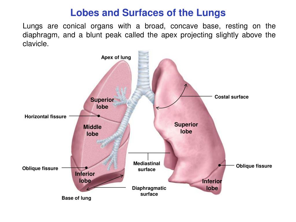
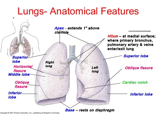
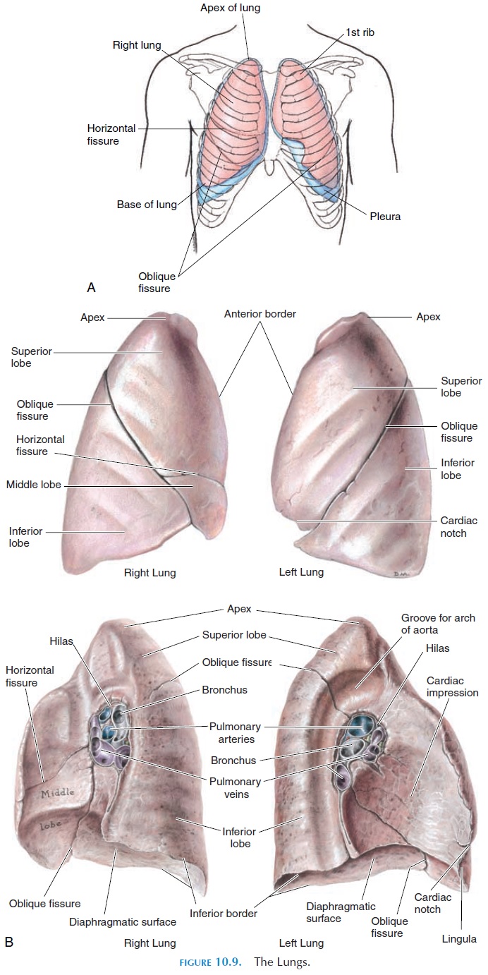
To fully grasp the significance of the lung apex, one must look at where it sits within the human body. Unlike the broad, concave base of the lung that rests directly on the diaphragm, the apex extends upward into the root of the neck.
Specifically, the apex of the lung rises above the level of the sternal end of the first rib, reaching approximately 2 to 4 centimeters above the level of the middle third of the clavicle (collarbone). Because of this elevated position, it actually enters the lower neck region, covered by a dense layer of deep fascia known as suprapleural membrane, or Sibson's fascia. This membrane protects the lung tissue from the muscular pressures of the neck and helps anchor the pleural cavity.
The apex is closely related to several critical vascular and neurological structures that pass through the thoracic outlet into the upper limbs. These include:
- The subclavian artery and vein
- The brachial plexus (a network of nerves supplying the arm)
- The sympathetic chain and the first thoracic ganglion
- The phrenic and vagus nerves
Relations and Surroundings
Because the apex of the lung extends so high into the thoracic inlet, it is bound by rigid anatomical structures. Posteriorly, it relates to the neck of the first rib. Medially, it lies close to large blood vessels originating from the aortic arch, such as the common carotid and subclavian arteries.
The pleura—a double-layered membrane surrounding the lungs—extends even higher than the lung tissue itself. This means the cervical pleura forms a protective dome over the apex of the lung, maintaining negative pressure and facilitating smooth respiratory movements without friction against the neck structures.
Clinical Significance and Pathology
The unique anatomical positioning of the apex makes it uniquely susceptible to specific medical conditions. Because of its proximity to nerves, blood vessels, and bone, pathologies occurring in this region present with distinct clinical signs.
Pancoast Tumors (Superior Sulcus Tumors)
Perhaps the most famous clinical condition associated with this area is the Pancoast tumor. This is a specialized type of non-small cell lung cancer that originates specifically in the apex of the lung. Because tumors in this location grow outward into the surrounding chest wall and thoracic inlet rather than filling the main airway, they often do not cause typical early lung cancer symptoms like a persistent cough.

Instead, a Pancoast tumor compresses nearby neural and vascular structures, leading to a constellation of symptoms known as Pancoast syndrome:
- Severe Shoulder and Arm Pain: Caused by the invasion of the lower roots of the brachial plexus (typically C8 and T1).
- Horner's Syndrome: Results from disruption of the cervical sympathetic chain, characterized by constricted pupils (miosis), drooping eyelid (ptosis), and decreased sweating on one side of the face (anhidrosis).
- Weakness and Atrophy: Muscle wasting in the hand muscles due to brachial plexus involvement.
- Edema: Swelling in the arm caused by compression of the subclavian vein.
Apical Pneumothorax
Another common clinical issue involving the apex is a spontaneous pneumothorax, commonly known as a collapsed lung. Tall, thin, young males are particularly prone to developing small fluid- or air-filled sacs called blebs on the surface of the lung, most frequently located at the lung apex.
If one of these blebs ruptures, air leaks into the pleural space, causing the lung to pull away from the chest wall. Because air naturally rises, a small pneumothorax is often first detected radiologically at the apex of the lung on a standard upright chest X-ray.
Tuberculosis and Apical Cavitation
Historically, the apex of the lung has been recognized as a primary site for the reactivation of pulmonary tuberculosis (TB). Mycobacterium tuberculosis bacteria thrive in environments with high oxygen tension. Because the upright human posture creates a ventilation-perfusion mismatch where ventilation and oxygenation are highest at the top of the lungs, the apex provides an ideal, highly oxygenated microenvironment for aerobic bacteria like tuberculosis to multiply and form cavities.
Imaging and Physical Examination
Physicians evaluate the apex of the lung during both physical exams and diagnostic imaging.
During a physical examination, percussion (tapping on the chest) over the supraclavicular fossa can reveal areas of abnormal dullness, which might indicate a mass, consolidation, or fluid accumulation at the apex. Auscultation (listening with a stethoscope) in this region helps clinicians detect abnormal breath sounds or localized crackles indicative of infection or structural disease.
Radiologically, interpreting a chest X-ray requires careful attention to the apical zones. Due to overlapping shadows from the clavicles and first ribs, small apical nodules or early-stage Pancoast tumors can sometimes be difficult to spot. Therefore, specialized views—such as an apical lordotic view or a computed tomography (CT) scan—are frequently utilized to obtain a clearer, unobstructed look at the apex of the lung.
Conclusion
Though it occupies a relatively small space at the top of the thoracic cavity, the apex of the lung plays a critical role in human respiratory anatomy. Its position extending into the base of the neck places it in close contact with major nerves and blood vessels, explaining why localized pathologies like Pancoast tumors, blebs, and tuberculosis infections produce such distinct and severe clinical symptoms. A thorough understanding of this upper pulmonary region remains vital for diagnosing complex thoracic and neurological conditions effectively.