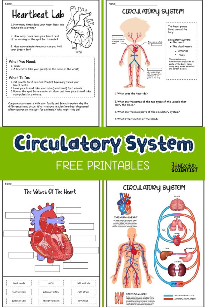
Cardiovascular System Worksheet: Full Study Guide
Mastering the circulatory network is one of the most fundamental milestones in any anatomy or biology curriculum. Whether you are preparing for an exam or designing a lesson plan, working through a structured worksheet cardiovascular system review is one of the most effective ways to convert abstract diagrams into lasting, functional knowledge.

This comprehensive study guide acts as both a self-contained learning module and a printable-ready resource. Below, you will explore the anatomy of the heart, trace the double-loop circulatory pathway, review the cellular components of blood, and test your retention with an interactive practice section complete with an answer key.
Core Anatomy: Structures of the Human Heart
The human heart is a muscular organ roughly the size of a closed fist, located in the mediastinum between the lungs. It functions as a dual pump operating simultaneously to power two distinct circulatory circuits.
The Four Chambers
- Right Atrium: Receives oxygen-depleted blood returning from the systemic circulation via the superior and inferior vena cava.
- Right Ventricle: Pumps this deoxygenated blood into the pulmonary artery toward the lungs for gas exchange.
- Left Atrium: Receives newly oxygenated blood returning from the lungs through the four pulmonary veins.
- Left Ventricle: Features the thickest myocardium of any chamber; it generates the high hydrostatic pressure needed to propel oxygenated blood through the aorta into the entire systemic circuit.
Heart Valves: Preventing Backflow
Valves maintain unidirectional blood flow through the heart. A standard anatomy worksheet will frequently ask you to categorize them into two groups:
- Atrioventricular (AV) Valves: Located between the atria and ventricles. The tricuspid valve sits on the right side, while the bicuspid (mitral) valve guards the left side. Both are anchored by chordae tendineae to papillary muscles, preventing prolapse during ventricular systole.
- Semilunar Valves: Located at the exits of the ventricles. The pulmonary valve sits at the base of the pulmonary trunk, and the aortic valve lies at the entrance to the ascending aorta.
The Circulatory Cycle: Step-by-Step Pathway
Understanding circulation requires following a single erythrocyte (red blood cell) as it navigates both loops of the cardiovascular system. Practice writing out this sequence until it becomes second nature:
- Deoxygenated blood arrives from peripheral tissues via the Superior and Inferior Vena Cava.
- It empties into the Right Atrium.
- Passes through the Tricuspid Valve into the Right Ventricle.
- Ejected past the Pulmonary Semilunar Valve into the Pulmonary Trunk and Pulmonary Arteries.
- Gas exchange occurs in the Capillary Beds of the Lungs (carbon dioxide is released, oxygen is bound to hemoglobin).
- Oxygen-rich blood returns via the Pulmonary Veins into the Left Atrium.
- Passes through the Bicuspid (Mitral) Valve into the Left Ventricle.
- Ejected past the Aortic Semilunar Valve into the Aorta.
- Distributed through systemic arteries, arterioles, capillaries (nutrient and gas exchange with tissues), venules, and veins back toward the vena cava.
Practice Worksheet Cardiovascular System Questions
Test your understanding with these practice exercises. Try completing them without referencing your notes before checking the answer key at the bottom.
Part 1: Multiple Choice
1. Which blood vessel carries oxygenated blood from the lungs back to the heart?
A) Pulmonary artery
B) Superior vena cava
C) Pulmonary vein
D) Aorta
2. Why is the myocardial wall of the left ventricle significantly thicker than that of the right ventricle?
A) It stores larger volumes of blood.
B) It must overcome greater peripheral resistance to pump blood throughout the entire body.
C) It pumps blood at a higher velocity directly into the cerebral circulation.
D) It protects the mitral valve from mechanical shear stress.
Video: Heart Structure & Functions Quiz | Human Circulatory System Quiz - Heart Chambers, Valves, & Vessels

3. Which component of blood is primarily responsible for hemostasis (clot formation)?
A) Erythrocytes
B) Leukocytes
C) Thrombocytes (platelets)
D) Albumin
Part 2: Matching Anatomy
Match the anatomical structure in Column A to its correct functional description in Column B.
- A. Sinoatrial (SA) Node — 1. Cord-like tendons connecting papillary muscles to AV valves
- B. Chordae Tendineae — 2. The primary biological pacemaker of the heart
- C. Tunica Media — 3. Smallest vessels where exchange of gases and nutrients occurs
- D. Capillaries — 4. Muscular, elastic middle layer of blood vessel walls
Part 3: Fill-in-the-Blank
1. The contraction phase of the cardiac cycle is known as __________, while the relaxation phase is called __________.
2. Blood vessels that carry blood away from the heart are categorized as __________, whereas vessels carrying blood toward the heart are __________.
3. The double-layered fibroserous sac that surrounds and protects the heart is the __________.
Answer Key and Explanations
Review your responses against the explanations below to pinpoint any areas requiring further review.
Part 1 Answers
- 1. C (Pulmonary vein): In the systemic circuit, veins carry deoxygenated blood and arteries carry oxygenated blood. However, in the pulmonary circuit, this rule reverses: pulmonary arteries carry deoxygenated blood to the lungs, and pulmonary veins return oxygenated blood to the left atrium.
- 2. B: The right ventricle only pumps blood a short distance into the low-resistance pulmonary circuit. The left ventricle must overcome systemic vascular resistance to circulate blood through every organ system, requiring a much thicker muscular layer.
- 3. C (Thrombocytes): Platelets aggregate at endothelial break sites and activate the coagulation cascade to prevent hemorrhage.
Part 2 Answers
- A matches 2: The Sinoatrial (SA) Node sits in the superior wall of the right atrium and initiates the electrical impulse driving heart rate.
- B matches 1: Chordae Tendineae tether the AV valve leaflets to prevent them from inverting into the atria during ventricular contraction.
- C matches 4: The Tunica Media consists of smooth muscle cells and elastic fibers, controlling vasodilation and vasoconstriction.
- D matches 3: Capillary walls consist of a single endothelial cell layer, facilitating rapid diffusion of respiratory gases, glucose, and metabolic wastes.
Part 3 Answers
- 1. Systole; Diastole
- 2. Arteries; Veins
- 3. Pericardium
Practical Study Tips for Cardiovascular Mastery
To retain this material over the long term, incorporate these three active recall strategies into your revision:
- Color-Code Flow Charts: Draw the heart from scratch using red ink for oxygenated structures (left side, pulmonary veins, aorta) and blue ink for deoxygenated pathways (right side, vena cava, pulmonary trunk). This visual contrast reinforces the two distinct circuits.
- Trace the Pulse Wave: Pair the mechanical events of the heart with its electrical conduction system (SA node → AV node → Bundle of His → Purkinje fibers). Understanding how electrical signaling coordinates chamber contraction makes ECG interpretation intuitive.
- Teach the Path: Recite the 9-step blood pathway out loud from memory without checking a diagram. Once you can articulate the route smoothly from the vena cava back to the aorta, you have mastered the foundational core of cardiovascular anatomy.