Interventions for Impaired Gas Exchange: Nursing Care
Impaired gas exchange is a critical nursing diagnosis defined as the excess or deficit in oxygenation and/or carbon dioxide elimination at the alveolar-capillary membrane. When this delicate physiological balance is disrupted, patients experience hypoxemia (low blood oxygen) or hypercapnia (excess carbon dioxide), leading to systemic complications. Implementing effective interventions for impaired gas exchange is vital for stabilizing patients, preventing respiratory failure, and restoring optimal tissue perfusion.

Understanding Impaired Gas Exchange
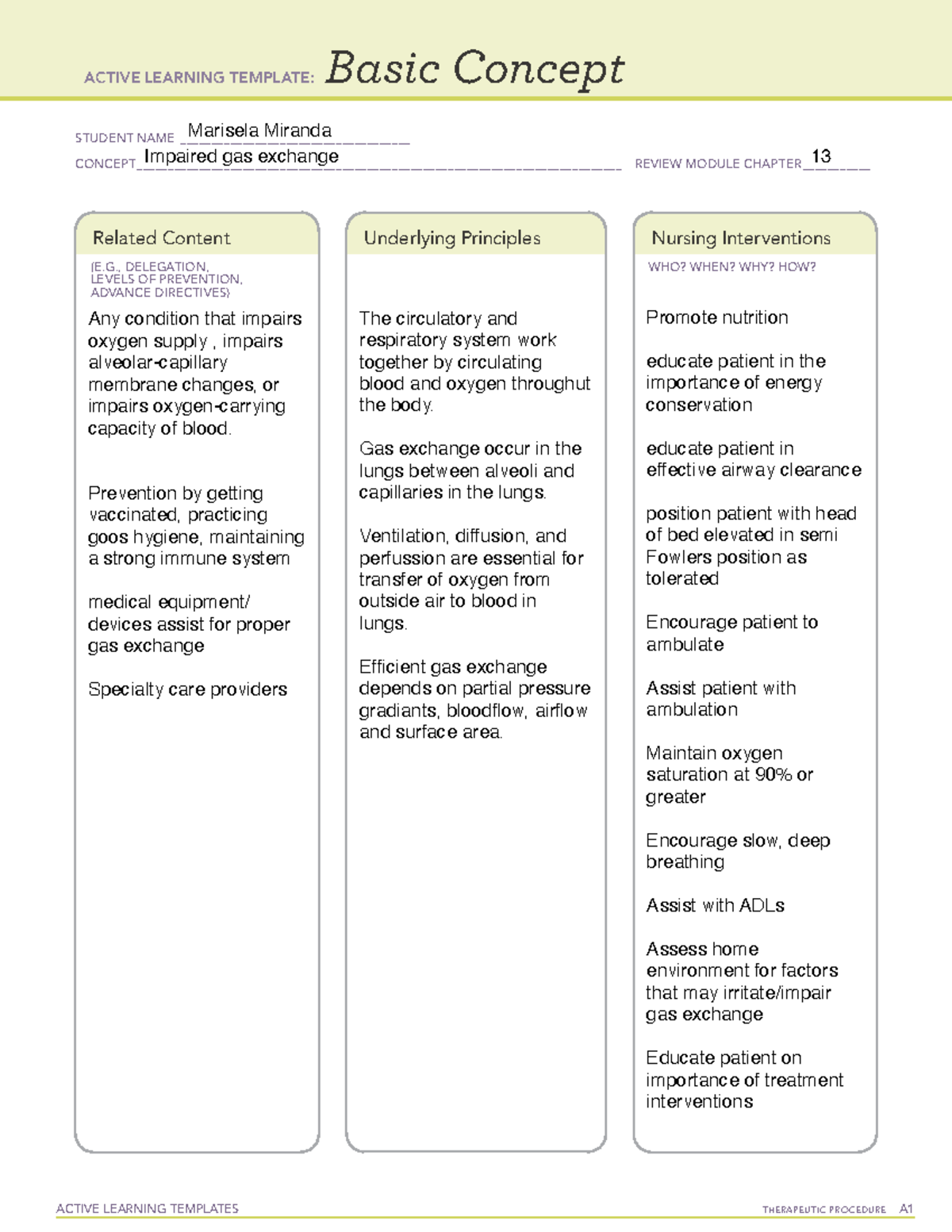
Before implementing specific therapies, clinicians must identify the root cause of the respiratory deficit. Gas exchange relies on three primary processes: ventilation (the movement of air in and out of the lungs), perfusion (blood flow through the pulmonary capillaries), and diffusion (the passive movement of gases across the alveolar-capillary membrane).
Conditions such as pneumonia, chronic obstructive pulmonary disease (COPD), pulmonary embolism, pulmonary edema, and acute respiratory distress syndrome (ARDS) frequently disrupt these processes. Recognizing early signs—such as tachypnea, dyspnea, restlessness, cyanosis, and abnormal arterial blood gas (ABG) values—allows for rapid, targeted treatment.
Patients with impaired gas exchange often exhibit characteristic clinical indicators that guide the urgency and scope of care:
- Abnormal arterial blood gases (e.g., low PaO2, high PaCO2)
- Tachypnea or irregular breathing patterns
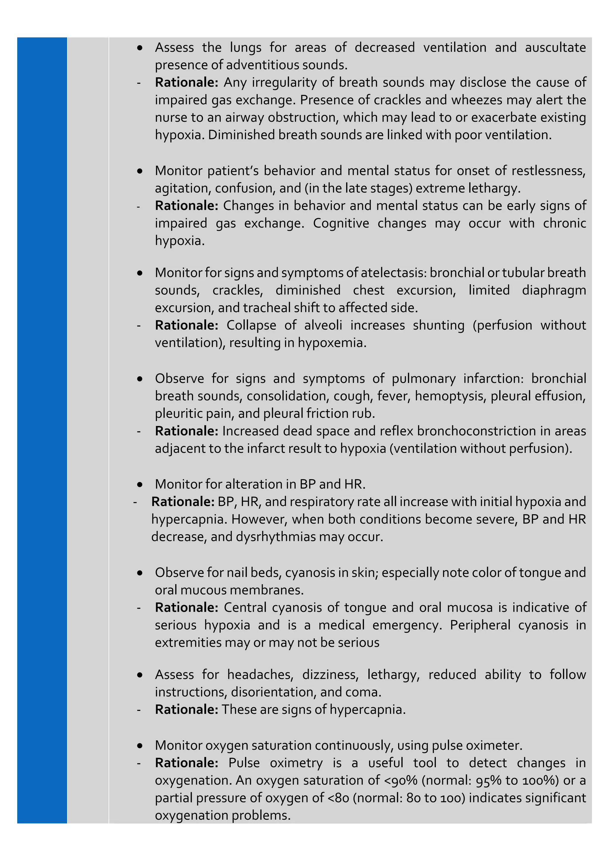
- Restlessness, confusion, or lethargy due to cerebral hypoxia
- Cyanosis (a late sign of hypoxia)
- Use of accessory muscles during respiration
Core Clinical Interventions for Impaired Gas Exchange
Managing compromised respiratory function requires a multi-faceted approach involving airway clearance, oxygen delivery, pharmacotherapy, and continuous monitoring. Below are the primary evidence-based interventions used in clinical settings.
1. Oxygen Therapy and Respiratory Support
Administering supplemental oxygen is the cornerstone of managing hypoxemia. The delivery method depends on the patient's specific needs and stability:
- Nasal Cannula: Ideal for patients requiring low-flow oxygen (1–6 L/min) for mild hypoxemia.
- Venturi Mask: Provides precise oxygen concentrations, making it especially useful for patients with chronic hypercapnic respiratory failure (such as those with COPD).
- Non-Rebreather Mask: Delivers high concentrations of oxygen (up to 90–100%) for acute, severe hypoxemia.
- High-Flow Nasal Cannula (HFNC): Delivers heated and humidified oxygen at high flow rates, washing out dead space in the upper airway and providing a mild positive end-expiratory pressure (PEEP) effect.
2. Positioning Strategies
Video: Core Concepts / Gas Exchange / Med-Surg Nursing

Patient positioning profoundly impacts ventilation-perfusion ($V/Q$) matching. Proper alignment can immediately improve oxygenation without altering medication or oxygen flow rates:
- High-Fowler’s or Semi-Fowler’s Position: Elevating the head of the bed to 45–90 degrees reduces abdominal pressure on the diaphragm, allowing for maximal chest expansion and improved lung volume.
- Good Lung Down: In unilateral lung diseases (such as pneumonia or a lung abscess), positioning the healthy lung in the dependent position improves perfusion to the better-ventilated alveoli, optimizing $V/Q$ matching.
- Prone Positioning: Frequently utilized in patients with severe ARDS, placing the patient face-down improves ventilation in the dorsal regions of the lungs and relieves pressure from the heart.
3. Airway Clearance Techniques
Retained pulmonary secretions obstruct airflow and worsen gas exchange. Clearing the airway is essential for effective ventilation:
- Controlled Coughing and Deep Breathing: Encouraging deep inspiration followed by a forceful cough helps mobilize secretions from smaller airways.
- Chest Physiotherapy (CPT): Includes postural drainage, percussion, and vibration to dislodge thick mucus plugs.
- Suctioning: Performed orally, nasopharyngeally, or endotracheally when the patient is unable to clear secretions independently. Care must be taken to pre-oxygenate the patient and limit suction passes to prevent hypoxia.
4. Pharmacological Management
Medications prescribed by the healthcare team directly target the underlying pathophysiology of respiratory impairment:
- Bronchodilators (Beta-agonists and Anticholinergics): Relax bronchial smooth muscle to relieve bronchospasms and open narrowed airways.
- Corticosteroids: Reduce airway inflammation and edema in conditions like asthma exacerbations and COPD.
- Diuretics: Eliminate excess fluid in patients experiencing pulmonary congestion secondary to heart failure or fluid overload.
- Mucolytics: Thin thick respiratory secretions, making them easier to expel.
Monitoring and Evaluation
Continuous assessment is necessary to gauge the effectiveness of implemented therapies and catch early signs of respiratory deterioration. Healthcare providers should prioritize the following monitoring parameters:
- Pulse Oximetry: Continuously monitor $SpO_2$ levels, keeping in mind target ranges prescribed for the specific patient (e.g., 88–92% for chronic CO2 retainers, 92–99% for standard patients).
- Arterial Blood Gases (ABGs): Review pH, $PaO_2$, $PaCO_2$, and bicarbonate levels to evaluate acid-base balance and the adequacy of ventilation and oxygenation.
- Respiratory Rate and Effort: Assess breathing frequency, depth, symmetry, and the use of accessory muscles.
- Auscultate lung sounds regularly to detect diminished breath sounds, crackles, wheezes, or rhonchi.
Patient Education and Lifestyle Modification
For long-term management, empowering the patient with knowledge is critical. Education should focus on adherence to prescribed inhaler regimens, recognizing early symptoms of a respiratory infection, practicing energy conservation techniques during daily activities, and smoking cessation programs. Adequate hydration and nutrition also support mucosal integrity and respiratory muscle strength, ensuring sustained recovery and prevention of future gas exchange deficits.