Urgent Frog Dissection Guide for Exams
Staring down a lab practical with zero preparation can induce instant panic. Whether you missed class or simply need a rapid refresher before stepping into the laboratory, this urgent frog dissection guide for exams is designed to get you up to speed in minutes. We will skip the fluff and focus strictly on what examiners demand: proper pin placement, safe incision techniques, and the identification of high-yield internal organs.

Pre-Lab Setup and External Anatomy
Before you make your first cut, examiners will often test your knowledge of external features. Place the preserved specimen dorsal side up in your dissecting pan and pin down all four legs securely.
Quickly locate and mentally note these high-frequency exam points:
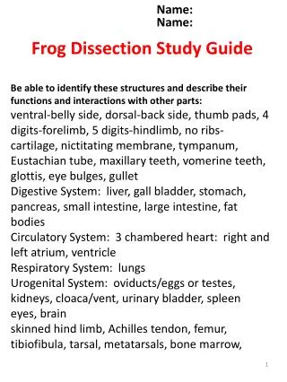
- Tympanic membrane: The circular disk behind each eye used for hearing.
- Nictitating membrane: The clear lower eyelid that protects the eye underwater.
- Digits: Note the webbed hind feet (for swimming) and unwebbed forelimbs (for support).
- Sexual dimorphism: Check the thumbs on the forelegs. If they are enlarged and swollen, it is a male (used for clasping females during mating).
Step-by-Step Incision Protocol
Speed and cleanliness dictate your practical score. Messy cuts obscure vital organs and lead to point deductions. Follow this precise cutting sequence to expose the body cavity without damaging underlying tissue.
- Lift the skin of the lower abdomen with forceps just above the pelvic bone.
- Make a small, horizontal snip with your scissors, then cut anteriorly (upward) along the midline all the way to the jaw.
- Make lateral cuts outward near the arms and legs to create flaps. Pin these flaps securely to the wax pan.
- Repeat this exact Y-shaped cutting procedure for the underlying muscle wall, lifting carefully as you cut to avoid puncturing the liver or intestines.
Urgent Frog Dissection Guide for Exams: Internal Organs to Memorize
Once the body cavity is pinned open, you have seconds to locate the structures your instructor is likely pointing a probe at. Scan for these major organ systems immediately.

The Digestive System
The digestive tract is usually the easiest to identify due to its sheer size and central location.
- Liver: The massive, dark brown, three-lobed organ dominating the upper chest cavity.
- Stomach: A large, J-shaped muscular sac located underneath the left lobe of the liver. Follow it to find the small intestine.
- Small Intestine: The coiled tube leading from the stomach, held together by a membrane called the mesentery.
- Gallbladder: A small, greenish sac tucked underneath the central lobe of the liver (frequently asked on practical tags).
- Pancreas: A small, yellowish gland nestled in the curve between the stomach and the first part of the small intestine.
Circulatory and Respiratory Systems
Examiners love testing heart anatomy and respiratory organs because they are immediately visible upon opening the chest.
- Heart: Located near the top of the body cavity. Look for the single thick-walled ventricle at the bottom and the two smaller atria at the top, encased in a thin sac called the pericardium.
- Lungs: Spongy, sac-like organs located dorsal to the heart on either side. Note that frog lungs are relatively simple compared to mammalian lungs.
- Spleen: A dark red, spherical organ attached to the mesentery near the lower portion of the small intestine, responsible for filtering blood.
Urogenital System
Depending on your grade level, reproductive and excretory organs wrap up most lab practical exams.
- Kidneys: Flat, dark red, elongated organs pressed against the dorsal (back) wall of the frog.
- Fat Bodies: Bright yellow, finger-like projections attached to the kidneys, used for storing energy during hibernation.
- Testes (Males): Bean-shaped, yellowish organs attached to the anterior end of the kidneys.
- Ovaries / Oviducts (Females): If your specimen is female, the body cavity may be packed with dark mass eggs or convoluted white-to-clear tubes (oviducts).
Last-Minute Exam Tips for Success
If you are minutes away from your practical test, keep these final pointers in mind to secure every possible point:
- Keep it moist: Use your dropper to periodically squirt water on the specimen. Dried-out tissue looks abnormal and obscures structures.
- Use the probe, not your fingers: Grading instructors deduct points for sloppy tearing. Use a blunt probe to gently push fat bodies or lobes of the liver aside to reveal hidden structures like the gallbladder or pancreas.
- Know your left from right: Remember that anatomical left and right are always from the specimen's perspective, not yours as you look down into the pan.